Genetic variants play a role in susceptibility to infectious diseases. Not everyone will get the norovirus or a particular strain of the flu — due to genetic variants. New research shows that genetics also plays a role in the severity of COVID-19.
Genetics and COVID-19: Who gets sick?
NOTE: This article is several years old and primarily based on studies on the initial Covid variants, not the currently circulating variants. It is included here for historical purposes.
Before we begin: Research on COVID-19 is being produced at a record pace, which is great. But not all publications are relevant or good sources of information. As software developers like to say: ‘garbage in, garbage out‘. With poor data and flawed models, many of the research studies being churned out on COVID-19 fall in the ‘garbage out’ category.
I’ve focused this article on quality research that has been peer-reviewed and published in legitimate journals, when possible. Anything referenced in a pre-print (non-peer-reviewed) publication will be clearly marked as such.
Genetics studies are just now beginning to come out on SARS-CoV-2 susceptibility, and I’m sure there will be many more to come! So consider this the beginning, not the final word, on genetics and SARS-CoV-2
Terminology:
Let’s define a few terms so that we are all on the same page:
- SARS-CoV-2 (severe acute respiratory syndrome coronavirus 2) is the name of the novel coronavirus identified by researchers in Wuhan, China in December 2019.
- COVID-19 is the name for the disease/symptoms caused by the SARS-CoV2 virus.
- Case: As everyone is probably aware, the definition of a COVID-19 ‘case’ is ever-changing and depends on the source. Numbers for cases differ according to each country’s definition, which also changes over time.[ref] This makes it difficult to make comparisons between countries and over time. Importantly, the varying definitions will impact the way that COVID-related genetic variants are identified.
Coronavirus Quick Facts:
- Since the 1960s, researchers have identified more than 50 different coronaviruses, most of which only cause diseases in other animals. There are now 7 known human coronaviruses.
- The viruses can be categorized as Alphacoronavirus or Betacoronavirus, depending on their structure. The common coronaviruses are one cause of the common cold.[ref]
- The SARS-CoV-2 virus is a betacoronavirus, an enveloped virus with a single strand of RNA. It enters human cells through the ACE2 receptor protein on the cell surface. Inside the cell, it uses the host’s cellular machinery to replicate.[ref]
- Previous serious viral outbreaks in the coronavirus-family include SARS-CoV and MERS-CoV.
- Coronaviruses that cause the common cold make rounds every year in a seasonal respiratory virus pattern.
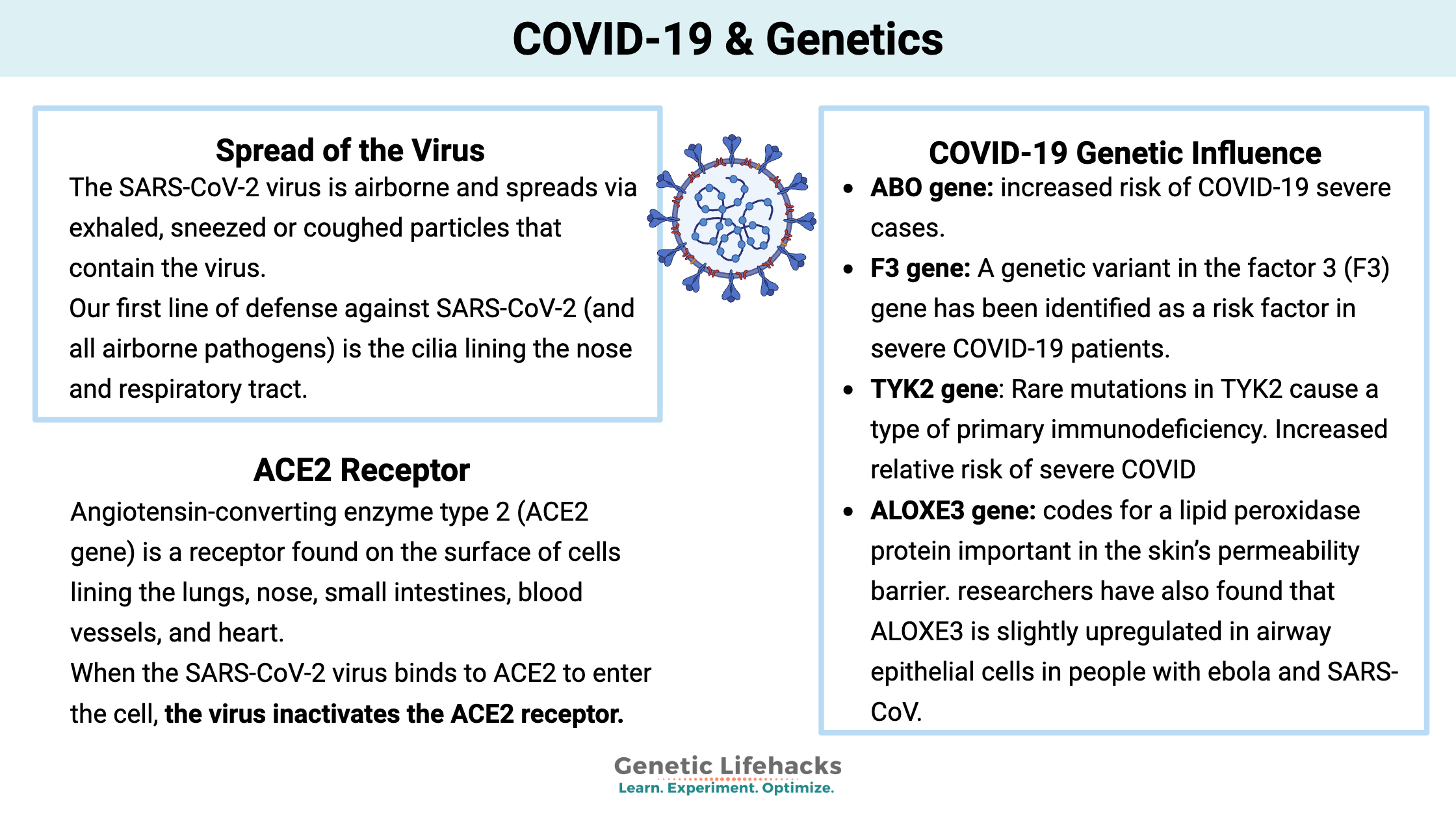
Spread of the virus:
The SARS-CoV-2 virus is airborne and spreads via exhaled, sneezed or coughed particles that contain the virus. Currently, almost all contact-tracing confirmed cases have spread in the home or in enclosed indoor environments that have poor air circulation. (In other words, outdoor spread or in well-ventilated spaces is not common.)[ref][pre-print ref]
Past coronavirus research clearly shows that cool, dry conditions facilitate the spread of the virus. This would include both winter-time weather as well as indoor, air-conditioned environments. Humid, warm environments decrease the spread (but only in areas where people don’t use the A/C when it gets hot). Studies in India show that SARS-CoV-2 spreads quickly in cities with a strong power infrastructure and A/C usage, but in crowded cities with little air conditioning, the virus does not seem to be spreading.[ref]
Asymptomatic cases:
Like most viruses, there is a huge variation in how sick people get with SARS-CoV-2. The majority of people are asymptomatic and don’t have symptoms. Of the people who do have symptoms (cases), the majority have cold-like symptoms. Unfortunately, a minority of people end up with severe symptoms that can lead to death, generally from either pneumonia or vascular complications.
The estimates on asymptomatic individuals range from initial studies showing 72% (Diamond Princess cruise ship) to initial serology studies showing 95% of people are asymptomatic or had symptoms mild enough not to be tested.[ref][pre-print ref] The CDC’s more recent estimates are that about half are asymptomatic or very mild, depending on which phone survey or serological study is used.[ref][ref]
Asymptomatic spread of the virus: While there is a lot of speculation about how often asymptomatic transmission of the virus occurs, research does show that asymptomatic carriers can spread the virus with close contact. One study looked at over 2000 close contacts of asymptomatic carriers and found the infection rate of close contacts with asymptomatic patients was 4%. [ref] It is likely that longer contact times allow more of a viral load to be transferred.
Pre-symptomatic spread is also well documented. Infected people can spread the virus a day or two before their symptoms begin.
The body’s first line of defense against respiratory viruses:
Our first line of defense against SARS-CoV-2 (and all airborne pathogens) is the cilia lining the nose and respiratory tract. The epithelial cells lining the upper respiratory tract can have as many as 200-300 cilia per cell, or little projections, on the surface. These cilia ‘beat’ or move back and forth in a coordinated fashion, moving pathogens and pollutants up and out of the airway. The mucous lining the surface collects the debris. The ciliary movement is thought to decrease in the elderly, and it is impaired by exposure to alcohol, cigarette smoke, and air pollution.[ref]
Nitric oxide is produced in the nose and helps to fight off pathogens. Researchers theorize that breathing through the nose rather than the mouth can decrease susceptibility.[ref]
ACE2 receptor:
What happens when the virus breaches the ‘first line of defense”? A virus can’t just wiggle its way into a cell. It must have a way of binding with a receptor on the cell surface and then bringing its RNA or DNA into the cell. Different viruses use different cell surface receptors to enter the cell.
- Angiotensin-converting enzyme type 2 (ACE2 gene) is a receptor found on the surface of cells lining the lungs, nose, small intestines, blood vessels, and heart.[ref]
- ACE2 is part of the renin-angiotensin-aldosterone system (RAAS), which controls blood pressure, fluid balance, and electrolytes in the body. ACE2 is considered to be an anti-inflammatory part of this system; it balances out the ACE1 enzyme.[ref]
- Researchers theorize that low levels of ACE2 (due to older age, hypertension, diabetes, cardiovascular disease) are likely to increase lung inflammation and coagulation.[ref]
- When the SARS-CoV-2 virus binds to ACE2 to enter the cell, the virus inactivates the ACE2 receptor. Researchers theorize that part of the hyperstimulation and cytokine storm produced by severe COVID-19 is due to diminished ACE2 production.[ref]
Some researchers theorize that inhibiting ACE1 will boost ACE2 and thus be beneficial in COVID-19 patients. ACE1 inhibitors often having coughing as a side effect, so angiotensin II receptor blockers (ARBs), such as the ‘-sartan’ blood pressure drugs, may be a better choice.[ref]
The research on whether more or less ACE2 is beneficial is still preliminary and not yet confirmed.
Antibodies, T-cell response, Complement System:
There are two main categories of the body’s immune system:
- Innate Immune Response: jumps into action immediately and fights off a variety of pathogens.
- Adaptive Immune Response: a more targeted approach that takes longer to initiate. The adaptive immune response can take days or weeks to develop a response to a specific pathogen. The advantage here is that the adaptive immune system creates a ‘memory’ of the pathogen and reacts immediately if exposed again.
With SARS-CoV-2 and other viruses, the innate immune response consists of interferon and other cytokines that can kill the cells containing the virus.
The adaptive immune response is unique to each virus and bacterium. In general, the adaptive immune response has two types of white blood cells:
- B cells (or B lymphocytes): created in the bone marrow and able to produce antibodies specific to an invader
- T cells (or T lymphocytes): maturing in the thymus and able to stimulate a variety of responses and long-term memory
Researchers are finding that while not everyone produces antibodies (from B cells) when they have COVID-19 (the majority do), some of the people who didn’t produce antibodies did produce T cells that fight and remember SARS-CoV-2.[pre-print]
Research from 2016 shows that T cell response was important in immunity for SARS-CoV and MERS-CoV, with antibodies fairly quickly disappearing for both of those viruses.[ref]
Not everyone produces antibodies to SARS-CoV2:
We hear a lot about antibody testing, but several recent studies show that some people who test positive for COVID-19 may not produce antibodies. The T-cell response may be more important for some people.
- A preprint released on June 28th found that 80% of COVID-19 patients had IgG antibodies (leaving 20% without).[pre-print]
- Antibodies may diminish very quickly in people who are asymptomatic. People with severe COVID-19 produced more antibodies than mild/moderate cases.[ref]
- German research shows that the majority of COVID patients created a T-cell response, even if they didn’t produce antibodies (12% didn’t). Additionally, the study found that about 80% of the control group (patients without COVID) had T-cells from previous coronavirus exposure that cross-reacted to SARS-CoV-2. This cross-reactivity may not provide complete immunity for everyone, but it may lessen symptoms.[pre-print] (Several previous studies have shown that about 40-50% of the population has cross-reactive T-cells, likely from previous coronavirus exposures.)
Complement system
The innate immune system also includes the ‘complement system’. The complement particles circulate at low levels all the time in the bloodstream, and, when activated, cause inflammation and call in phagocytes to kill off pathogens.
The complement system is considered to be the oldest part of our immune system by evolutionary biologists. It is thought to be one of the first systems that evolved to identify bacteria as being different from the host.[ref]
There are several ways that the complement system can be activated. At low levels, it is always on patrol for pathogens – or anything that is not ‘self’ (known as the alternative pathway). Part of the complement system can enhance the immune response when antibodies are already present for the pathogen (known as the classical pathway). Finally, a part of the complement system is called mannose-binding lectin, which can bind to mannose on pathogens, marking the pathogen for destruction. Additionally, complement activation can increase clotting.
The complement system is composed of many different molecular components – some that recognize pathogens, some that cause activation of the immune response, and others that keep the complement system in check. Too much complement activation can result in the destruction of some of the body’s cells, such as happens in the eyes of elderly people with age-related macular degeneration. Another example of overactivation of the complement system in young people is atypical hemolytic uremic syndrome, which causes damage to the endothelial cells lining the blood vessels.[ref]
An August 2020 paper in the journal Nature Medicine, explains that one big difference with severe COVID-19 patients may lie in activation, or rather, overactivation, of the complement system. The research was conducted using nasal swabs from patients at NYC hospitals and using genetic data from the UK Biobank.
The researchers found that people with age-related macular degeneration, a condition caused by a dysregulated complement system, were at a much higher risk of dying from COVID-19. Yes, the researchers took into account age and sex when determining whether age-related macular degeneration (ARMD) was a risk factor.
Additionally, the researchers found that people who were genetically more likely to have coagulation disorders (thrombosis, thrombocytopenia) were also more likely to have severe consequences from COVID-19. For example, the researchers identified an SNP in the factor 3 gene, which codes for part of the coagulation cascade, as increasing the risk of COVID-19 mortality by 2-fold. (listed in the genetics section below)
One more interesting study may add even more evidence to the idea that the complement system dysregulation is at the root of severe COVID-19. The study by Stanford researchers found that the immune response goes awry with severe COVID-19. What was interesting (in terms of activating the complement system) was the following quote:
“The scientists also found elevated levels of bacterial debris, such as bacterial DNA and cell-wall materials, in the blood of those COVID-19 patients with severe cases. The more debris, the sicker the patient — and the more pro-inflammatory substances circulating in his or her blood. The findings suggest that in cases of severe COVID-19, bacterial products ordinarily present only in places such as the gut, lungs, and throat may make their way into the bloodstream, kick-starting enhanced inflammation that is conveyed to all points via the circulatory system.”
Genetic studies on Covid (as of 2022):
Genome-Wide Association Study from Italy and Spain (blood type, innate immune system):
A recent study published in the New England Journal of Medicine looked at the genetic data of almost 2,000 patients in Italy and Spain who had severe respiratory failure with COVID-19. The researchers used a method known as a genome-wide association study (GWAS) to statistically determine whether people with severe COVID-19 had any genetic differences from a control group. This GWAS found two statistically significant spots in the genome.
One hotspot in the genome was in the ABO blood group. The study found that in the ABO gene, a marker identifying type A blood was statistically linked to severe COVID-19. The researchers then further divided the patients by blood type and found that people with type A blood were more likely to have a severe course with COVID-19, while people with type O blood were the least likely to have severe COVID-19.
- Type A: 45% increased relative risk of severe COVID
- Type O: 35% decreased relative risk of severe COVID
So what does it really mean to be at a 45% increased relative risk?
Example (with made-up numbers) to illustrate relative risk:
Let’s say that the average risk of severe COVID-19 is 10% (10 out of 100) for a 70-year-old man with type 2 diabetes who comes down with the SARS-CoV-2 virus… If that person had type A blood, his risk is 45% higher than a similar person without type A blood. Thus, his risk would rise to 14.5% (or 14.5 out of 100). Similarly, if his blood type were type O, his risk would decrease by 35%, and thus the severe COVID risk would be 7%.
The genetic variant used in the study (listed below) links to blood type for most people, but not all. It gets a bit complicated and takes multiple SNPs to define blood type for everyone. So if you know your blood type already, just go with that as being more accurate than the SNP below in the genotype report.
Chromosome 3 region:
The second ‘hotspot’ in the genome-wide association study was a genetic variant in a region of chromosome 3 that could change the amount of a couple of immune system molecules the body produces. This region of chromosome 3 is not a specific gene, per se. The researchers found the “risk allele … is associated with reduced expression of CXCR6 and increased expression of SLC6A20, and LZTFL1 is strongly expressed in human lung cells …”. CXCR6 (chemokine receptor type 6) is a protein expressed in T cells.
Note that the risk allele listed in the study is rs11385942. This SNP is not included in 23andMe or AncestryDNA, but another SNP, rs10490770, is inherited together with the original SNP for over 99% of people. So we will use rs10490770 as a proxy here.
Coagulation factor and Complement system genes (Nature Medicine paper)[ref]:
F3 gene: A genetic variant in the factor 3 (F3) gene has been identified as a risk factor in severe COVID-19 patients. Factor 3 is part of the coagulation cascade and linked with fibrinogen levels.
Meta-analysis of genetics studies:
A July 2021 meta-analysis of genetics studies on COVID-19 attempted to consolidate what is known about genetics and who gets sick. Many of the genetics studies included in the analysis point to the same variants (listed above). The new meta-analysis, though, shed additional light on a variant in the TYK2 gene, including an alternative SNP that is found in 23andMe data.
TYK2 gene: encodes a tyrosine kinase that is important in the immune system. It is part of the interferon signaling pathway, which is activated to fight off viral infections. Rare mutations in TYK2 cause a type of primary immunodeficiency.
Neanderthals, Genetics, and COVID:
An interesting pre-print points out that the risk factor listed in the NEJM study (above) is found in an area of chromosome 3 inherited from Neanderthals. Getting even more specific, the area of chromosome 3 is likely more related to Croatian Neanderthals than to Siberian Neanderthals.[ref]
Let me explain a bit about who carries Neanderthal genetic data. (If you have your results from 23andMe, there is a Neanderthal Ancestry report.)
Quick Neanderthal History: Scientists have found multiple skeletons from several different species of ancient humans, including Neanderthals and Denisovans. Over the last decade, researchers have sequenced the genomes of the Neanderthals and Denisovans from their skeletal remains. This genetic research shows how some Homo sapiens (modern humans) interbred with Neanderthals and Denisovans. The Neanderthals mainly inhabited Europe and Asia and were not found in Africa.[ref]
Essentially, people who are of African ancestry do not have Neanderthal or Denisovan DNA mixed in with the modern human DNA. People of East Asian and European ancestry are likely to have around 2% of their DNA from Neanderthals.
Why is this important? This link with a region of chromosome 3 inherited from Neanderthals could explain statistical differences in why some population groups (European, Asian) are hit harder by COVID-19 than others.
UK Biobank Study:
A pre-print study published July 6th looked at the genetic variation between the genomes of 180 people who died of COVID-19 in comparison with a control group of over 1000 people. The study found that mutations in 4 genes were statistically significant. These uncommon mutations are found in less than 1% of the population but recorded at a number disproportionately higher in the people who died compared with controls.
Why this study is interesting: While a relatively small study, the results point to rare variants in specific immune-related genes rather than more common SNPs being important. While rare variants are just that (rare) on a population level, pretty much everyone carries some rare mutations. For example, if there are a handful of mutations (found in a little less than 1% of the population) that are important for COVID-19 mortality, adding those rare mutations together statistically makes sense for a severe case fatality rate of 1-3%.
BRF2 gene: The first mutation (rs138763430) is in the BRF2 gene. This gene codes for a transcription factor important in RNA polymerase II binding. Viruses hijack our own cells’ RNA polymerase to replicate, so it is likely that this gene is important in viral replication.
ERAP2 gene: Another mutation (rs150892504 ) found in the ERAP2 gene codes for a zinc metalloaminopeptidase protein. This protein is important for the way that antigens are presented via the HLA class 1 binding peptides. Antigen presentation then allows for T cells to respond and kill the infected cells.
ALOXE3 gene: codes for a lipid peroxidase protein important in the skin’s permeability barrier. Other mutations in ALOXE3 cause a condition where the skin is red, dry, and scaly with increased infection risk (ichthyosis). While more well researched for its role in the skin and liver, researchers have also found that ALOXE3 is slightly upregulated in airway epithelial cells in people with Ebola and SARS-CoV. [pre-print][ref][ref]
In addition to the mutation listed below, a second ALOXE3 mutation (rs151256885) not covered by 23andMe or AncestryDNA data was also found more frequently in patients who died of COVID-19.[preprint]
APOE E4 allele:
A recent study found that people with the APOE E4/E4 genotype are at a 2-fold higher risk of mortality from COVID-19. People with the E4 allele were also at a slightly higher overall risk of testing positive for COVID-19.[ref]
The E4/E4 genotype is linked with a greatly increased risk of Alzheimer’s disease. (Check your genetic data here, if you want to know)
Which genetic variants are likely to be ruled out?
We can learn quite a bit from studies that show that theoretically important variants are not significant.
- ACE I/D: Recent studies show that the ACE Insertion/Deletion has not been shown to impact SARS-CoV.[ref] Initial studies, though, showed a possible correlation at the population level. Keep in mind that each country defined cases differently and used different testing methods, so it is possible that the correlation is not meaningful.[ref][ref] (Read more about ACE I/D and check your genes)
- HFE, VDR: One study ruled out the HFE mutations that cause hemochromatosis as well as the vitamin D related variants.[ref]
- ACE2 and TMPRSS2: Many studies theorize that the ACE2 genetic variants should impact COVID-19, but none have clearly shown a big statistical link so far in cases. Some genetic testing companies initially jumped to a conclusion that variants linked to increased ACE2 or TMPRSS2 would cause severe COVID-19, but the research doesn’t show this. In fact, for ACE2, the research seems to show the opposite, with ACE2 playing an anti-inflammatory role.[ref][ref][ref][ref]
- Alternatively, it could be that tissue-specific ACE2 levels are important. Some research points to increased ACE2 in the lung tissue of elderly patients with severe COVID-19.[pre-print] ACE2 levels are likely important in COVID-19, but there are many additional lifestyle factors that change ACE2 levels other than genetic variants.
Severe COVID-19 Genotype Report:
Access this content:
An active subscription is required to access this content.
Lifehacks for Boosting Immune Function:
A robust innate immune response (that initial response to a pathogen) is needed to clear the SARS-CoV-2 virus.
Below are a few of the research-backed ways to help stack the odds in your favor with the SARS-CoV-2 virus. These ‘lifehacks’ may help increase your immune response — but please know there are many variables, known and unknown, at play in whether you have symptoms with SARS-CoV-2.
No magic pills here… just support to keep your immune function in excellent condition.
Circadian rhythm and immune function:
Your immune system is better equipped to fight off pathogens during the day. This makes sense when you think about how society interacted before electricity — generally, people went to bed rather than going out on the town.
Animal studies show that the time of day matters a lot in viral replication. Mice infected with a virus right before the rest phase (e.g., bedtime) had a 10-fold greater viral replication than the mice infected at the start of their active phase. Additionally, mice with a core circadian rhythm gene knocked out have a higher viral replication load, day or night.[ref]
The best thing for healthy people to prevent COVID-19 symptoms may end up being to avoid social activities at night and go to bed at a reasonable hour.
Jet lag also messes with your circadian rhythm and increases your susceptibility to viruses. Animal studies on jet lag clearly show increased susceptibility to viral bronchitis and increased replication of the virus.[ref] A similar circadian disruption occurs when people stay up later than usual, exposed to bright lights — or are switching over to working nights (such as healthcare workers). Read more about circadian rhythm and immune function.
Vitamin D:
Quite a few epidemiological studies have pointed out the link between vitamin D deficiency and susceptibility to more severe COVID-19.[ref][ref]
A vitamin D blood test is your best bet for knowing your levels, and spending some time out in the sun should help to boost your levels if needed.
What about vitamin D as a supplement? A recent cell line study tested a variety of known drugs and other compounds against SARS-CoV2. The study found several known prescription drugs (with major side effects), as well as calcitriol, a synthetic form of vitamin D3, was possibly effective against SARS-CoV2. Calcitriol stood out as being effective in several different types of cells, including nasal epithelial cells. Plus, it is inexpensive and safe.[pre-print ref]
Melatonin:
Released at night from the pineal gland, melatonin is vital to the immune system. Research studies on sepsis show that melatonin may be beneficial for reducing excessive cytokine production and restoring mitochondrial function. Many animal studies show that melatonin enhances immune function prior to stimulation, and it may also tamp down an excessive immune system response.[ref][ref][ref][ref]
You can boost your own body’s production of melatonin by eliminating exposure to light in the blue wavelengths at night. Light at night (at 480nm, blue light) triggers a receptor in the retina, causing melatonin production to be dampened. Either turn off electronics/lights or wear blue light-blocking glasses. Both will increase your endogenous melatonin production.
Will supplemental melatonin help? Quite a few researchers are suggesting that melatonin supplements are a good idea for combating COVID. Some suggest that supplemental melatonin be used as a prophylaxis by everyone to prevent COVID-19.[ref][ref] There is currently a clinical trial underway to determine the effectiveness of supplemental melatonin.[ref]
Dual histamine blockers (more severe cases):
Cetirizine plus famotidine: The hyperactive inflammatory response in severe COVID-19 patients was battled with cetirizine and famotidine in a hospital-based study. The results showed that the percentage of patients on ventilators and the mortality rate was lower for the patients (n=110) on the dual histamine blockade… but there was no official placebo group for comparison.[pre-print ref]
Why would this work? Blocking the histamine response should reduce the overactive inflammatory cascade. Histamine, a signaling molecule, is released by mast cells in response to pathogens (or allergens), but histamine also acts as a signal to trigger other mast cells to degranulate – thus worsening the overall inflammatory cascade.
Recap of your genes:
| Gene | RS ID | Your Genotype | Notes for Your Genotype | Effect allele | Effect allele frequency |
|---|---|---|---|---|---|
| ABO | rs657152 | — | typicalincrease risk of COVID-19 severityincreased risk of COVID-19 severe cases (along with other risk factors such as age and co-morbidities) | A | 0.37 |
| ABO | rs10490770 | — | no increased riskincreased relative risk for severe COVID-19increased relative risk for severe COVID-19; greater increase in relative risk of severe COVID for patients under age 60 vs. over age 60 | C | 0.07 |
| F3 | rs72729504 | — | typical~2-fold increased risk of severe COVID-19 outcome~2-fold increased risk of severe COVID-19 outcome | T | 0.019 |
| intergenic | rs45574833 | — | typicalincreased risk of hospitalization with COVID-19increased risk of hospitalization with COVID-19 | A | 0.014 |
| intergenic | rs12064775 | — | typicalincreased risk of hospitalization with COVID-19increased risk of hospitalization with COVID-19 | G | 0.01 |
| TYK2 | rs34536443 | — | typicalincreased relative risk of severe COVID-19, decreased TYK2increased relative risk of severe COVID-19, decreased TYK2 | C | 0.03 |
| TMEM181 | rs117665206 | — | typicalmutation found more frequently in COVID-19 patients who died[ pre-print ]mutation found more frequently in COVID-19 patients who died[ pre-print ] | T | 0.006 |
| ALOXE3 | rs147149459 | — | typicalmutation found more frequently in patients who died[ pre-print ]mutation found more frequently in COVID-19 patients who died[ pre-print ] | A | 0.001 |
Related Articles and Topics:
Boosting NAD+ levels to fight the diseases of aging:
Explore the research about how nicotinamide riboside (NR) and NMN are being used to reverse aging. Learn about how your genes naturally affect your NAD+ levels, and how this interacts with the aging process.
Fisetin: Anti-aging senolytic and powerful antioxidant
Recent research on the flavonoid fisetin shows that it may be a potent supplement for improving health in aging. Fisetin has been shown to act as a senolytic, clearing out senescent cells in animal and cells studies.
Anxiety: Genetic variants that increase the risk of anxiety disorders
This article covers genetic variants related to anxiety disorders. This is a big topic, and new research is coming out all the time. The information here is presented for educational/informational purposes. This means… learn all that you can about the physiological and genetic reasons for anxiety disorders, but do talk with your doctor before making any medical decisions.
Originally published July 10, 2020. Updated August 2020.
Works Cited:
Actual Coronavirus Infections Vastly Undercounted, C.D.C. Data Shows – The New York Times. https://www.nytimes.com/2020/06/27/health/coronavirus-antibodies-asymptomatic.html.
Ahmad, Tauseef, et al. “COVID-19: Zoonotic Aspects.” Travel Medicine and Infectious Disease, Feb. 2020, p. 101607. ScienceDirect, doi:10.1016/j.tmaid.2020.101607.
Al-Sadeq, Duaa W., and Gheyath K. Nasrallah. “The Incidence of the Novel Coronavirus SARS-CoV-2 among Asymptomatic Patients: A Systematic Review.” International Journal of Infectious Diseases, July 2020. ScienceDirect, doi:10.1016/j.ijid.2020.06.098.
—. “The Incidence of the Novel Coronavirus SARS-CoV-2 among Asymptomatic Patients: A Systematic Review.” International Journal of Infectious Diseases, July 2020. ScienceDirect, doi:10.1016/j.ijid.2020.06.098.
Alsufyani, Hadeel A., and James R. Docherty. “The Renin Angiotensin Aldosterone System and COVID-19.” Saudi Pharmaceutical Journal, July 2020. ScienceDirect, doi:10.1016/j.jsps.2020.06.019.
Anderson, George, and Russel J. Reiter. “Melatonin: Roles in Influenza, Covid‐19, and Other Viral Infections.” Reviews in Medical Virology, vol. 30, no. 3, May 2020. PubMed Central, doi:10.1002/rmv.2109.
Asselta, Rosanna, et al. ACE2 and TMPRSS2 Variants and Expression as Candidates to Sex and Country Differences in COVID-19 Severity in Italy. SSRN Scholarly Paper, ID 3559608, Social Science Research Network, 21 Mar. 2020. papers.ssrn.com, doi:10.2139/ssrn.3559608.
Carrillo-Vico, Antonio, et al. “Melatonin: Buffering the Immune System.” International Journal of Molecular Sciences, vol. 14, no. 4, Apr. 2013, pp. 8638–83. PubMed Central, doi:10.3390/ijms14048638.
CDC. “Coronavirus Disease 2019 (COVID-19).” Centers for Disease Control and Prevention, 11 Feb. 2020. www.cdc.gov, https://www.cdc.gov/coronavirus/2019-ncov/cases-updates/commercial-lab-surveys.html.
Clinical and Immunological Assessment of Asymptomatic SARS-CoV-2 Infections | Nature Medicine. https://www.nature.com/articles/s41591-020-0965-6.
Colunga Biancatelli, Ruben Manuel Luciano, et al. “Melatonin for the Treatment of Sepsis: The Scientific Rationale.” Journal of Thoracic Disease, vol. 12, no. Suppl 1, Feb. 2020, pp. S54–65. PubMed, doi:10.21037/jtd.2019.12.85.
Could Nasal Nitric Oxide Help to Mitigate the Severity of COVID-19? https://www.ncbi.nlm.nih.gov/pmc/articles/PMC7200356/.
Delanghe, Joris R., Marijn M. Speeckaert, and Marc L. De Buyzere. “COVID-19 Infections Are Also Affected by Human ACE1 D/I Polymorphism.” Clinical Chemistry and Laboratory Medicine (CCLM), vol. 58, no. 7, De Gruyter, June 2020, pp. 1125–26. www.degruyter.com, doi:10.1515/cclm-2020-0425.
Delanghe, Joris R., Marijn M. Speeckaert, and Marc L. De Buyzere. “The Host’s Angiotensin-Converting Enzyme Polymorphism May Explain Epidemiological Findings in COVID-19 Infections.” Clinica Chimica Acta, vol. 505, June 2020, pp. 192–93. ScienceDirect, doi:10.1016/j.cca.2020.03.031.
Edgar, Rachel S., et al. “Cell Autonomous Regulation of Herpes and Influenza Virus Infection by the Circadian Clock.” Proceedings of the National Academy of Sciences, National Academy of Sciences, Aug. 2016. www.pnas.org, doi:10.1073/pnas.1601895113.
Ehlers, Anna, et al. “BMAL1 LINKS THE CIRCADIAN CLOCK TO VIRAL AIRWAY PATHOLOGY AND ASTHMA PHENOTYPES.” Mucosal Immunology, vol. 11, no. 1, Jan. 2018, pp. 97–111. PubMed Central, doi:10.1038/mi.2017.24.
Ellinghaus, David, et al. “Genomewide Association Study of Severe Covid-19 with Respiratory Failure.” New England Journal of Medicine, vol. 0, no. 0, Massachusetts Medical Society, June 2020, p. null. Taylor and Francis+NEJM, doi:10.1056/NEJMoa2020283.
García, Irene García, et al. “A Randomized Multicenter Clinical Trial to Evaluate the Efficacy of Melatonin in the Prophylaxis of SARS-CoV-2 Infection in High-Risk Contacts (MeCOVID Trial): A Structured Summary of a Study Protocol for a Randomised Controlled Trial.” Trials, vol. 21, no. 1, June 2020, p. 466. PubMed, doi:10.1186/s13063-020-04436-6.
Grant, William B., et al. “Evidence That Vitamin D Supplementation Could Reduce Risk of Influenza and COVID-19 Infections and Deaths.” Nutrients, vol. 12, no. 4, Apr. 2020. PubMed, doi:10.3390/nu12040988.
Hoffmann, Markus, et al. “SARS-CoV-2 Cell Entry Depends on ACE2 and TMPRSS2 and Is Blocked by a Clinically Proven Protease Inhibitor.” Cell, vol. 181, no. 2, Apr. 2020, pp. 271-280.e8. ScienceDirect, doi:10.1016/j.cell.2020.02.052.
Indoor Transmission of SARS-CoV-2 | MedRxiv. https://www.medrxiv.org/content/10.1101/2020.04.04.20053058v1.
Klein et al. – 2020 – Sex, Age, and Hospitalization Drive Antibody Respo.Pdf. https://www.medrxiv.org/content/10.1101/2020.06.26.20139063v1.full.pdf. Accessed 15 July 2020.
Klein, Sabra, et al. Sex, Age, and Hospitalization Drive Antibody Responses in a COVID-19 Convalescent Plasma Donor Population. preprint, Infectious Diseases (except HIV/AIDS), 28 June 2020. DOI.org (Crossref), doi:10.1101/2020.06.26.20139063.
Kuo et al. – ApoE E4e4 Genotype and Mortality with COVID-19 in .Pdf. https://www.medrxiv.org/content/10.1101/2020.06.19.20134908v1.full.pdf.
Liu, William J., et al. “T-Cell Immunity of SARS-CoV: Implications for Vaccine Development against MERS-CoV.” Antiviral Research, vol. 137, Jan. 2017, pp. 82–92. PubMed Central, doi:10.1016/j.antiviral.2016.11.006.
Lippi, Giuseppe, et al. “Do Genetic Polymorphisms in Angiotensin Converting Enzyme 2 (ACE2) Gene Play a Role in Coronavirus Disease 2019 (COVID-19)?” Clinical Chemistry and Laboratory Medicine (CCLM), vol. 1, no. ahead-of-print, June 2020. www.degruyter.com, doi:10.1515/cclm-2020-0727.
Lu, Chang, et al. Genetic Risk Factors for Death with SARS-CoV-2 from the UK Biobank. preprint, Epidemiology, 5 July 2020. DOI.org (Crossref), doi:10.1101/2020.07.01.20144592.
Lund Håheim, Lise. “Epithelial Cilia Is the First Line of Defence against Coronavirus; Addressing the Observed Age-Gradient in the COVID-19 Infection.” Medical Hypotheses, vol. 143, Oct. 2020, p. 110064. ScienceDirect, doi:10.1016/j.mehy.2020.110064.
Martel, Jan, et al. “Could Nasal Nitric Oxide Help to Mitigate the Severity of COVID-19?” Microbes and Infection, vol. 22, no. 4, 2020, pp. 168–71. PubMed Central, doi:10.1016/j.micinf.2020.05.002.
Meltzer, David O., et al. “Association of Vitamin D Deficiency and Treatment with COVID-19 Incidence.” MedRxiv: The Preprint Server for Health Sciences, May 2020. PubMed, doi:10.1101/2020.05.08.20095893.
Reference, Genetics Home. “ALOXE3 Gene.” Genetics Home Reference. ghr.nlm.nih.gov, https://ghr.nlm.nih.gov/gene/ALOXE3.
Reiter, Russel J., et al. “Therapeutic Algorithm for Use of Melatonin in Patients With COVID-19.” Frontiers in Medicine, vol. 7, May 2020. PubMed Central, doi:10.3389/fmed.2020.00226.
Srinivasan, Venkataramanujam, et al. “Melatonin in Bacterial and Viral Infections with Focus on Sepsis: A Review.” Recent Patents on Endocrine, Metabolic & Immune Drug Discovery, vol. 6, no. 1, Jan. 2012, pp. 30–39. PubMed, doi:10.2174/187221412799015317.
Tsang, Tim K., et al. “Effect of Changing Case Definitions for COVID-19 on the Epidemic Curve and Transmission Parameters in Mainland China: A Modelling Study.” The Lancet Public Health, vol. 5, no. 5, Elsevier, May 2020, pp. e289–96. www.thelancet.com, doi:10.1016/S2468-2667(20)30089-X.
V, Antony Aroul Raj, et al. “The Contribution of Dry Indoor Built Environment on the Spread of Coronavirus: Data from Various Indian States.” Sustainable Cities and Society, July 2020, p. 102371. ScienceDirect, doi:10.1016/j.scs.2020.102371.
Verdecchia, Paolo, et al. “The Pivotal Link between ACE2 Deficiency and SARS-CoV-2 Infection.” European Journal of Internal Medicine, vol. 76, 2020, pp. 14–20. PubMed, doi:10.1016/j.ejim.2020.04.037.
Vernot, Benjamin, et al. “Excavating Neandertal and Denisovan DNA from the Genomes of Melanesian Individuals.” Science (New York, N.Y.), vol. 352, no. 6282, Apr. 2016, pp. 235–39. PubMed Central, doi:10.1126/science.aad9416.