Genes that Impact Ferritin Levels

Your body relies on iron as a way to carry oxygen — it is vital and yet only needed in limited amounts. Serum ferritin levels are one way of measuring the amount of iron in your body, and altered lab results can indicate an underlying problem.

This article explains ferritin test levels, genetic variants that impact ferritin, and how to increase ferritin with supplements or foods Members will see their genotype report below, plus additional solutions in the Lifehacks section. Join today 

What is ferritin?

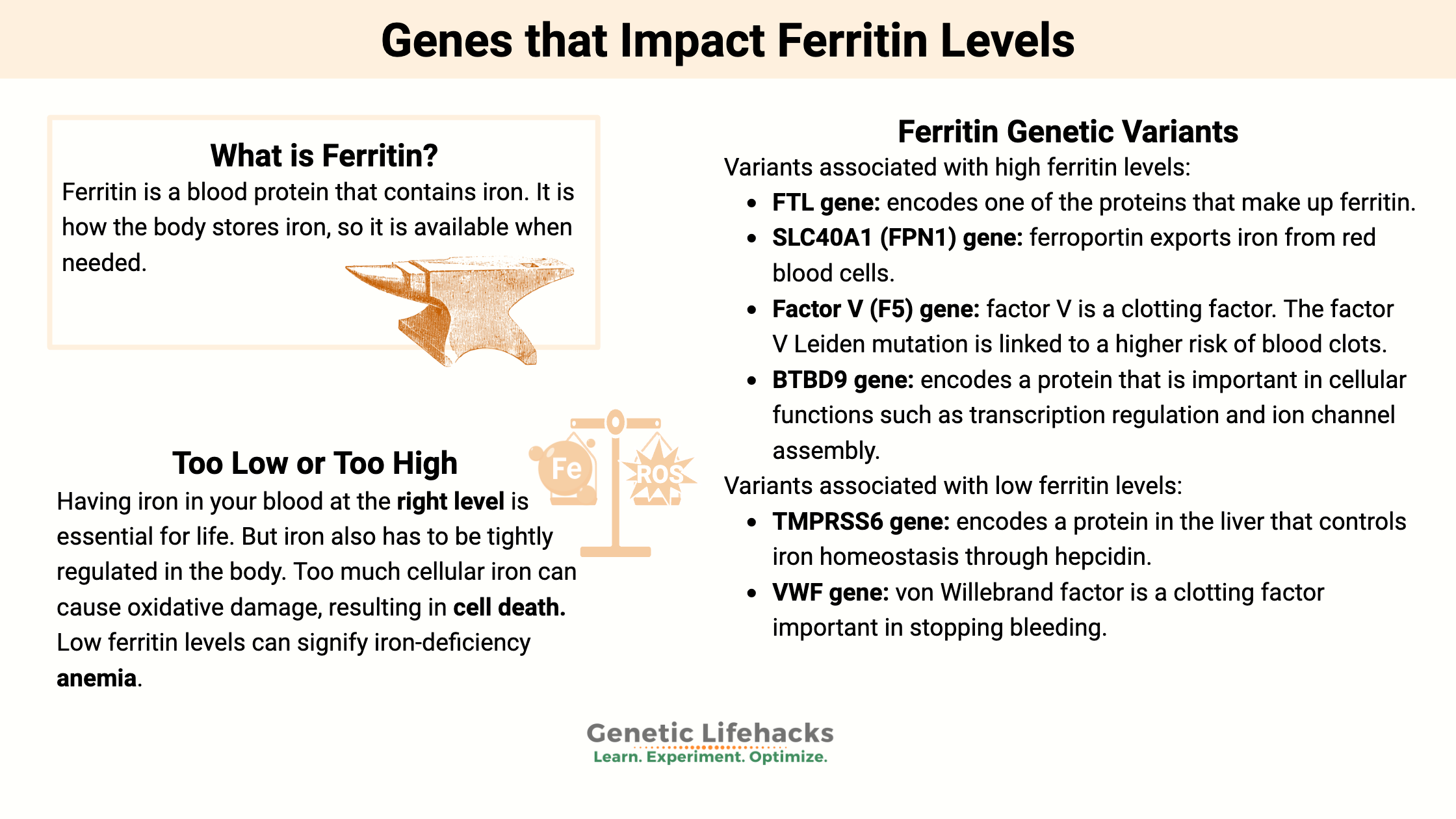
Ferritin is a blood protein that contains iron. It is how the body stores iron, so it is available when needed.

Having iron in your blood at the right level is essential for life. But iron also has to be tightly regulated in the body. Too much cellular iron can cause oxidative damage, resulting in cell death.[ref]

Iron is also essential for many pathogens – bacteria, malaria, etc. Thus, your body regulates iron as a way to keep pathogens from being able to survive.[ref]

Ferritin, the iron storage protein, is primarily found in the liver, bone marrow, muscles, and spleen. About 70-80% of your iron is stored as ferritin when you are healthy.[ref] This iron storage gives the body access to an adequate supply whenever needed.

Each ferritin protein is made up of subunits that come together to form the structure. Within this structure, iron is deposited. “A single ferritin molecule of this type can hold up to 4300 iron ions in its central cavity”. In addition to iron, ferritin also holds phosphate.[ref]

Ferritin blood tests: normal serum ranges

According to the Mayo Clinic, the normal range for a ferritin test is:[ref]

  • For men, 24 to 336 mg/l (or ng/mL)
  • For women, 11 to 307 mg/l

You may notice that the normal range varies a bit depending on the testing company. For example, Mount Sinai has the normal range as 12 -300 ng/mL for men and 12 – 150 ng/mL.

Low or high ferritin levels:

Low ferritin levels can signify iron-deficiency anemia, but it is also found in other conditions. Your doctor will likely do additional tests to determine the cause for you. For example, testing for transferrin saturation and serum iron levels can shed light on the root cause.

According to the CDC, iron status is defined using the following criteria:[ref]

Iron Status Stored Iron (ferritin) Transport iron (transferrin saturation) Functional iron (serum iron
iron-deficiency anemia low low low
iron-deficiency erythropoiesis low low normal
iron depletion low normal normal
iron overload high high normal

Causes of low ferritin:

Ferritin is the storage protein for iron in the body, and low iron levels lead to low ferritin levels.[ref]

Blood loss or low iron intake are the leading causes of low ferritin levels in healthy people. Other causes can include inflammatory bowel disease or celiac disease, which decreases iron absorption.[ref]

Symptoms of iron deficiency anemia include:[ref]

  • fatigue or weakness
  • pale skin
  • fast heartbeat, shortness of breath
  • headache, dizziness, lightheadedness
  • cold hands and feet
  • inflammation or soreness of your tongue (burning tongue, cracks)
  • pica (craving for eating dirt, ice)

Causes of high ferritin:

Several diseases cause higher ferritin levels.

  • Hemochromatosis, a genetic reason for iron build-up (check your genes here)
  • Porphyria — A group of disorders caused by an enzyme deficiency that affects your nervous system and skin
  • Inflammatory disorders such as Rheumatoid arthritis
  • Liver disease
  • Hyperthyroidism
  • Blood cancers (leukemia and Hodgkin’s lymphoma)

High ferritin levels can also be caused by:

  • Multiple blood transfusions
  • Alcohol abuse
  • Excessive supplemental iron

Inflammation usually causes ferritin levels to rise. Inflammatory cytokines, such as IL-1β, IL-6, and TNFα, increase the creation of the ferritin molecule. Excess ROS, such as hydrogen peroxide, can also increase ferritin.[ref]

Chronically elevated inflammatory cytokines, such as is seen in periodontitis (gum disease), are associated with higher serum ferritin levels. Higher ferritin levels are also found, on average, in people with diabetes.[ref]

Proteins that regulate iron:

When looking at the genetic variants that link to higher ferritin levels, we must go beyond just the genes that encode the ferritin proteins. Instead, the main genetic drivers of higher ferritin averages include other proteins involved in regulating iron levels.

Here’s a quick overview:

Ferroportin is the protein responsible for transporting iron out of cells. It is found in the small intestines, liver cells, and macrophages (a white blood cell type). For example, in the small intestines, it regulates the transport of iron from food into the bloodstream. Ferroportin is an important regulator when there is too much iron available, inflammation, or a lot of red blood cells being produced.[ref]

Hepcidin is another iron regulatory protein. It regulates how much iron can circulate by inhibiting ferroportin. In this way, hepcidin essentially regulates how much iron is absorbed in the intestines and allowed into circulation. During times of acute inflammation, hepcidin levels rise, causing iron levels to fall.[ref]

Transferrin is a glycoprotein that transports iron in the blood plasma. The TF gene encodes transferrin, and it is produced in the liver. Transferrin binds to iron (usually in the intestines) and transports it until it encounters a transferrin receptor (on red blood cells). It binds with the transferrin receptor and releases iron ions.

Transferrin levels decrease in inflammation, cancer, and some diseases (acute-phase protein that decreases instead of increasing)


Ferritin Genotype Report:

Members: Log in to see your data below.
Not a member? Join here. Membership lets you see your data right in each article and also gives you access to the member’s only information in the Lifehacks sections.

There are over 300 genes that impact iron levels, in one way or another, in humans and almost all other living organisms. Iron regulation is complex and touches on so many processes in the body.[ref]

Genetics is only part of the picture with ferritin (and iron). Infection and diet are also really important here. I’m going to cover the well-researched genetic variants that impact ferritin levels – and are found in your genetic raw data file. It means that there are likely other variants or mutations that also impact ferritin that aren’t covered here… You can’t rule anything out with this information.

Men and women have differences in the need for iron and how iron is handled in the body. Some of the genetic studies take this into account, and I’ve noted it below if the study results apply to one biological sex and not the other. The same goes for different ancestry groups. Unfortunately, not all studies take into account population ancestry or sex.

FTL gene: encodes one of the proteins that make up ferritin. (Note that there are not any common variants that seem to have an impact here. Ferritin is completely essential to the regulation of iron and life.)

Check your genetic data for rs104894685 (AncestryDNA):

  • A/G: carrier of a rare mutation related to ferritin (double-check this with a clinical-grade test)[ref]
  • G/G: typical

Members: Your genotype for rs104894685 is .

Check your genetic data for rs397514540 (AncestryDNA):

  • C/T: carrier of a rare mutation related to high ferritin (double-check this with a clinical-grade test)[ref]
  • C/C: typical

Members: Your genotype for rs397514540 is .

SLC40A1 (FPN1) gene: ferroportin exports iron from red blood cells.

Check your genetic data for rs11568350 (AncestryDNA):

  • C/C: typical
  • A/C: higher ferritin levels (African American men)[ref]
  • A/A: higher ferritin levels (African American men)[ref]; children less likely to be anemic[ref]

Members: Your genotype for rs11568350 is .

TF gene: encodes transferrin protein

Check your genetic data for rs1799852 (23andMe v5; AncestryDNA):

  • C/C: typical
  • C/T: lower serum transferrin levels, slightly higher ferritin levels (on average)
  • T/T: lower serum transferrin levels, slightly higher ferritin levels (on average)[ref]

Members: Your genotype for rs1799852 is .

Check your genetic data for rs3811647 (23andMe v4, v5; AncestryDNA):

  • A/A: higher ferritin (relative to G/G)[ref]
  • A/G: higher ferritin (relative to G/G)
  • G/G: typical

Members: Your genotype for rs3811647 is .

TMPRSS6 gene: encodes a protein in the liver that controls iron homeostasis through hepcidin

Check your genetic data for rs855791 (23andMe v4, v5):

  • G/G: typical
  • A/G: lower ferritin levels (Caucasian men)
  • A/A: lower ferritin levels (Caucasian men)[ref]

Members: Your genotype for rs855791 is .

SLC17A1 gene: encodes a phosphate transporter

Check your genetic data for rs17342717 (23andMe v4, v5; AncestryDNA):

  • C/C: typical
  • C/T: higher ferritin (check to see if you have a HFE mutation related to hemochromatosis)
  • T/T: higher ferritin (check to see if you have a HFE mutation related to hemochromatosis)[ref]

Members: Your genotype for rs17342717 is .

BTBD9 gene: encodes a protein that is important in cellular functions such as transcription regulation and ion channel assembly.

Check your genetic data for rs9296249 (23andMe v4, v5):

  • C/C: higher serum ferritin levels[ref]
  • C/T: higher serum ferritin levels
  • T/T: typical (most common genotype)

Members: Your genotype for rs9296249 is .

Check your genetic data for rs3923809 (23andMe v4, v5; AncestryDNA):

  • A/A: increased risk of restless leg[ref][ref]; serum ferritin levels decreased 26%[ref] (most common genotype in Caucasians, minor allele in Asian populations)
  • A/G: serum ferritin levels decreased 13%
  • G/G: typical risk of RLS

Members: Your genotype for rs3923809 is .

VWF gene: von Willebrand factor is a clotting factor important in stopping bleeding

Check your genetic data for rs1800386 (AncestryDNA):

  • C/T: reduced ferritin levels (women only)[ref] (may also indicate VWF deficiency and increased menstrual bleeding)
  • T/T: typical

Members: Your genotype for rs1800386 is .

Factor V (F5) gene: factor V is a clotting factor. The factor V Leiden mutation is linked to a higher risk of blood clots. This variation is also linked to increased ferritin levels in premenopausal women.

Check your genetic data for rs6025 (23andMe v4, v5; AncestryDNA):

  • C/C: typical
  • C/T: one copy of factor V Leiden (read this article); increased ferritin levels in women (due to decreased menstrual bleeding)
  • T/T: two copies of factor V Leiden (read this article); increased ferritin levels in women (due to decreased menstrual bleeding)[ref]

Members: Your genotype for rs6025 is .


Lifehacks:

Always get a blood test to determine your iron levels before supplementing. Iron is essential but not benign. You definitely don’t want to overdo it with supplemental iron. Talk with a doctor or pharmacist if you have any questions about iron, especially regarding supplemental iron interactions with other medications.

According to the CDC, adults need at least 8 mg of iron per day. Premenopausal women need around 18mg/day, depending on blood loss due to menstruation.

How can you increase ferritin levels?

If you are low in iron, there are several dietary changes that may help to increase your iron levels.

Foods high in iron include[ref]:

Food Milligrams/serving Percent DV*
Breakfast cereals, fortified with 100% of the DV for iron, 1 serving 18 100
Oysters, eastern, cooked with moist heat, 3 ounces 8 44
White beans, canned, 1 cup 8 44
Chocolate, dark, 45%–69% cacao solids, 3 ounces 7 39
Beef liver, pan-fried, 3 ounces 5 28
Lentils, boiled and drained, ½ cup 3 17
Spinach, boiled and drained, ½ cup 3 17
Tofu, firm, ½ cup 3 17
Kidney beans, canned, ½ cup 2 11
Sardines, Atlantic, canned in oil, drained solids with bone, 3 ounces 2 11
Chickpeas, boiled and drained, ½ cup 2 11
Tomatoes, canned, stewed, ½ cup 2 11
Beef, braised bottom round, trimmed to 1/8” fat, 3 ounces 2 11
Potato, baked, flesh and skin, 1 medium potato 2 11
Cashew nuts, oil roasted, 1 ounce (18 nuts) 2 11
Green peas, boiled, ½ cup 1 6
Chicken, roasted, meat and skin, 3 ounces 1 6
Rice, white, long-grain, enriched, parboiled, drained, ½ cup 1 6
Bread, whole wheat, 1 slice 1 6
Bread, white, 1 slice 1 6
Raisins, seedless, ¼ cup 1 6

The rest of this article is for Genetic Lifehacks members only.  Consider joining today to see the rest of this article.

Food and Supplement Interactions with iron:

Member Content:

An active subscription is required to access this content.

Join Here for full access to this article, genotype reports, and much more!


Already a member? Log in below.


Related Articles and Topics:

Specialized Pro-resolving Mediators: Getting Rid of Chronic Inflammation
Chronic inflammation is at the root of all diseases. New research discusses how pro-resolving mediators are the key to the resolution of inflammation.

Always tired? Genetic reasons for fatigue
Are you always tired even when you know you slept well? Discover more about the newest research on fatigue and how genetic susceptibility plays a part in some people.

Elevated Fibrinogen: Risk factor for blood clots
Fibrinogen is a protein that is essential for creating blood clots when you get a wound. But higher levels of fibrinogen are a major risk factor for heart disease and DVT. Learn how your genes impact your fibrinogen level.

Building Up Iron: Hemochromatosis mutations
A couple of common mutations can cause you to build up iron, leading to iron overload or hemochromatosis. This is one genetic disease where knowledge is really powerful – you can completely prevent hemochromatosis through blood donations.

References:

Bell, Steven, et al. “A Genome-Wide Meta-Analysis Yields 46 New Loci Associating with Biomarkers of Iron Homeostasis.” Communications Biology, vol. 4, no. 1, Feb. 2021, pp. 1–14. www.nature.com, https://doi.org/10.1038/s42003-020-01575-z.

Benyamin, Beben, et al. “Variants in TF and HFE Explain ∼40% of Genetic Variation in Serum-Transferrin Levels.” American Journal of Human Genetics, vol. 84, no. 1, Jan. 2009, pp. 60–65. PubMed Central, https://doi.org/10.1016/j.ajhg.2008.11.011.

Bradley, Justin M., et al. “Ferritins: Furnishing Proteins with Iron.” Journal of Biological Inorganic Chemistry, vol. 21, 2016, pp. 13–28. PubMed Central, https://doi.org/10.1007/s00775-016-1336-0.

Chin, Dawn, et al. “Curcumin May Impair Iron Status When Fed to Mice for Six Months.” Redox Biology, vol. 2, Jan. 2014, pp. 563–69. ScienceDirect, https://doi.org/10.1016/j.redox.2014.01.018.

Ferritin Test – Mayo Clinic. https://www.mayoclinic.org/tests-procedures/ferritin-test/about/pac-20384928. Accessed 14 Feb. 2022.

Fischer, Jordie AJ, et al. “Is Untargeted Iron Supplementation Harmful When Iron Deficiency Is Not the Major Cause of Anaemia? Study Protocol for a Double-Blind, Randomised Controlled Trial among Non-Pregnant Cambodian Women.” BMJ Open, vol. 10, no. 8, Aug. 2020, p. e037232. PubMed Central, https://doi.org/10.1136/bmjopen-2020-037232.

Gasche, C., et al. “Iron, Anaemia, and Inflammatory Bowel Diseases.” Gut, vol. 53, no. 8, Aug. 2004, pp. 1190–97. PubMed Central, https://doi.org/10.1136/gut.2003.035758.

Guo, Lin-Na, et al. “Serum and Salivary Ferritin and Hepcidin Levels in Patients with Chronic Periodontitis and Type 2 Diabetes Mellitus.” BMC Oral Health, vol. 18, Apr. 2018, p. 63. PubMed Central, https://doi.org/10.1186/s12903-018-0524-4.

“Iron Deficiency Anemia – Symptoms and Causes.” Mayo Clinic, https://www.mayoclinic.org/diseases-conditions/iron-deficiency-anemia/symptoms-causes/syc-20355034. Accessed 14 Feb. 2022.

Kannengiesser, C., et al. “A New Missense Mutation in the L Ferritin Coding Sequence Associated with Elevated Levels of Glycosylated Ferritin in Serum and Absence of Iron Overload.” Haematologica, vol. 94, no. 3, Mar. 2009, pp. 335–39. DOI.org (Crossref), https://doi.org/10.3324/haematol.2008.000125.

McCullough, Kayla, and Subhashini Bolisetty. “Ferritins in Kidney Disease.” Seminars in Nephrology, vol. 40, no. 2, Mar. 2020, pp. 160–72. PubMed Central, https://doi.org/10.1016/j.semnephrol.2020.01.007.

Milman, Nils, et al. “Ferrous Bisglycinate 25 Mg Iron Is as Effective as Ferrous Sulfate 50 Mg Iron in the Prophylaxis of Iron Deficiency and Anemia during Pregnancy in a Randomized Trial.” Journal of Perinatal Medicine, vol. 42, no. 2, Mar. 2014, pp. 197–206. PubMed, https://doi.org/10.1515/jpm-2013-0153.

Moore, Hyatt, et al. “Periodic Leg Movements during Sleep Are Associated with Polymorphisms in BTBD9, TOX3/BC034767, MEIS1, MAP2K5/SKOR1, and PTPRD.” Sleep, vol. 37, no. 9, Sept. 2014, pp. 1535–42. PubMed Central, https://doi.org/10.5665/sleep.4006.

Muriuki, John Muthii, et al. “The Ferroportin Q248H Mutation Protects from Anemia, but Not Malaria or Bacteremia.” Science Advances, vol. 5, no. 9, Sept. 2019, p. eaaw0109. PubMed Central, https://doi.org/10.1126/sciadv.aaw0109.

NM_000146.4(FTL):C.286G>A (p.Ala96Thr) AND Neuroferritinopathy – ClinVar – NCBI. https://www.ncbi.nlm.nih.gov/clinvar/77958001/. Accessed 14 Feb. 2022.

Office of Dietary Supplements – Iron. https://ods.od.nih.gov/factsheets/Iron-HealthProfessional/. Accessed 14 Feb. 2022.

Pichler, Irene, et al. “Identification of a Common Variant in the TFR2 Gene Implicated in the Physiological Regulation of Serum Iron Levels.” Human Molecular Genetics, vol. 20, no. 6, Mar. 2011, pp. 1232–40. PubMed Central, https://doi.org/10.1093/hmg/ddq552.

Recommendations to Prevent and Control Iron Deficiency in the United States. https://www.cdc.gov/mmwr/preview/mmwrhtml/00051880.htm. Accessed 14 Feb. 2022.

Rivers, Charles A., et al. “Association of Ferroportin Q248H Polymorphism with Elevated Levels of Serum Ferritin in African-Americans in the Hemochromatosis and Iron Overload Screening (HEIRS) Study.” Blood Cells, Molecules & Diseases, vol. 38, no. 3, 2007, pp. 247–52. PubMed Central, https://doi.org/10.1016/j.bcmd.2006.12.002.

Sørensen, Erik, Katrine Grau, et al. “A Genetic Risk Factor for Low Serum Ferritin Levels in Danish Blood Donors.” Transfusion, vol. 52, no. 12, Dec. 2012, pp. 2585–89. PubMed, https://doi.org/10.1111/j.1537-2995.2012.03629.x.

Sørensen, Erik, Andreas S. Rigas, et al. “Genetic Factors Influencing Ferritin Levels in 14,126 Blood Donors: Results from the Danish Blood Donor Study.” Transfusion, vol. 56, no. 3, Mar. 2016, pp. 622–27. PubMed, https://doi.org/10.1111/trf.13397.

Stefansson, Hreinn, et al. “A Genetic Risk Factor for Periodic Limb Movements in Sleep.” The New England Journal of Medicine, vol. 357, no. 7, Aug. 2007, pp. 639–47. PubMed, https://doi.org/10.1056/NEJMoa072743.

Truman-Rosentsvit, Marianna, et al. “Ferritin Is Secreted via 2 Distinct Nonclassical Vesicular Pathways.” Blood, vol. 131, no. 3, Jan. 2018, pp. 342–52. PubMed Central, https://doi.org/10.1182/blood-2017-02-768580.
.


About the Author:
Debbie Moon is the founder of Genetic Lifehacks. Fascinated by the connections between genes, diet, and health, her goal is to help you understand how to apply genetics to your diet and lifestyle decisions. Debbie has a BS in engineering and also an MSc in biological sciences from Clemson University. Debbie combines an engineering mindset with a biological systems approach to help you understand how genetic differences impact your optimal health.

Find your next article: